Introduction to the Skeletal System
Humans are vertebrates, animals having a vertabral column or backbone. They rely on a sturdy internal frame that is centered on a prominent spine. The human skeletal system consists of bones, cartilage, ligaments and tendons and accounts for about 20 percent of the body weight.The living bones in our bodies use oxygen and give off waste products in metabolism. They contain active
tissues that consume nutrients, require a blood supply and change shape or remodel in response to variations in mechanical stress.
Bones provide a rigid framework, known as the skeleton, that support and protect the soft organs of the body.
The skeleton supports the body against the pull of gravity. The large bones of the lower limbs support the trunk when standing.
The skeleton also protects the soft body parts. The fused bones of the cranium surround the brain to make it less vulnerable to injury. Vertebrae surround and protect the spinal cord and bones of the rib cage help protect the heart and lungs of the thorax.
Bones work together with muscles as simple mechanical lever systems to produce body movement.
Bones contain more calcium than any other organ. The intercellular matrix of bone contains large amounts of calcium salts, the most important being calcium phosphate.
When blood calcium levels decrease below normal, calcium is released from the bones so that there will be an adequate supply for metabolic needs. When blood calcium levels are increased, the excess calcium is stored in the bone matrix. The dynamic process of releasing and storing calcium goes on almost continuously.
Hematopoiesis, the formation of blood cells, mostly takes place in the red marrow of the bones.
In infants, red marrow is found in the bone cavities. With age, it is largely replaced by yellow marrow for fat storage. In adults, red marrow is limited to the spongy bone in the skull, ribs, sternum, clavicles, vertebrae and pelvis. Red marrow functions in the formation of red blood cells, white blood cells and blood platelets.
Structure of Bone Tissue
There are two types of bone tissue: compact and spongy. The names imply that the two types differ in density, or how tightly the tissue is packed together. There are three types of cells that contribute to bone homeostasis. Osteoblasts are bone-forming cell, osteoclasts resorb or break down bone, and osteocytes are mature bone cells. An equilibrium between osteoblasts and osteoclasts maintains bone tissue.Compact Bone
Compact bone consists of closely packed osteons or haversian systems. The osteon consists of a central canal called the osteonic (haversian) canal, which is surrounded by concentric rings (lamellae) of matrix. Between the rings of matrix, the bone cells (osteocytes) are located in spaces called lacunae. Small channels (canaliculi) radiate from the lacunae to the osteonic (haversian) canal to provide passageways through the hard matrix. In compact bone, the haversian systems are packed tightly together to form what appears to be a solid mass. The osteonic canals contain blood vessels that are parallel to the long axis of the bone. These blood vessels interconnect, by way of perforating canals, with vessels on the surface of the bone.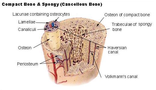
Spongy (Cancellous) Bone
Spongy (cancellous) bone is lighter and less dense than compact bone. Spongy bone consists of plates (trabeculae) and bars of bone adjacent to small, irregular cavities that contain red bone marrow. The canaliculi connect to the adjacent cavities, instead of a central haversian canal, to receive their blood supply. It may appear that the trabeculae are arranged in a haphazard manner, but they are organized to provide maximum strength similar to braces that are used to support a building. The trabeculae of spongy bone follow the lines of stress and can realign if the direction of stress changes.Bone Development & Growth
The terms osteogenesis and ossification are often used synonymously to indicate the process of bone formation. Parts of the skeleton form during the first few weeks after conception. By the end of the eighth week after conception, the skeletal pattern is formed in cartilage and connective tissue membranes and ossification begins.Bone development continues throughout adulthood. Even after adult stature is attained, bone development continues for repair of fractures and for remodeling to meet changing lifestyles. Osteoblasts, osteocytes and osteoclasts are the three cell types involved in the development, growth and remodeling of bones. Osteoblasts are bone-forming cells, osteocytes are mature bone cells and osteoclasts break down and reabsorb bone.
There are two types of ossification: intramembranous and endochondral.
Intramembranous
Intramembranous ossification involves the replacement of sheet-like connective tissue membranes with bony tissue. Bones formed in this manner are called intramembranous bones. They include certain flat bones of the skull and some of the irregular bones. The future bones are first formed as connective tissue membranes. Osteoblasts migrate to the membranes and deposit bony matrix around themselves. When the osteoblasts are surrounded by matrix they are called osteocytes.Endochondral Ossification
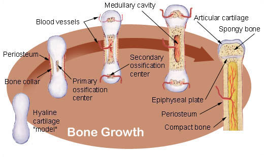
Endochondral ossification involves the replacement of hyaline cartilage with bony tissue. Most of the bones of the skeleton are formed in this manner. These bones are called endochondral bones. In this process, the future bones are first formed as hyaline cartilage models. During the third month after conception, the perichondrium that surrounds the hyaline cartilage "models" becomes infiltrated with blood vessels and osteoblasts and changes into a periosteum. The osteoblasts form a collar of compact bone around the diaphysis. At the same time, the cartilage in the center of the diaphysis begins to disintegrate. Osteoblasts penetrate the disintegrating cartilage and replace it with spongy bone. This forms a primary ossification center. Ossification continues from this center toward the ends of the bones. After spongy bone is formed in the diaphysis, osteoclasts break down the newly formed bone to open up the medullary cavity.The cartilage in the epiphyses continues to grow so the developing bone increases in length. Later, usually after birth, secondary ossification centers form in the epiphyses. Ossification in the epiphyses is similar to that in the diaphysis except that the spongy bone is retained instead of being broken down to form a medullary cavity.
When secondary ossification is complete, the hyaline cartilage is totally replaced by bone except in two areas. A region of hyaline cartilage remains over the surface of the epiphysis as the articular cartilage and another area of cartilage remains between the epiphysis and diaphysis. This is the epiphyseal plate or growth region.
Bone Growth
Bones grow in length at the epiphyseal plate by a process that is similar to endochondral ossification. The cartilage in the region of the epiphyseal plate next to the epiphysis continues to grow by mitosis.The chondrocytes, in the region next to the diaphysis, age and degenerate. Osteoblasts move in and ossify the matrix to form bone. This process continues throughout childhood and the adolescent years until the cartilage growth slows and finally stops.
When cartilage growth ceases, usually in the early twenties, the epiphyseal plate completely ossifies so that only a thin epiphyseal line remains and the bones can no longer grow in length. Bone growth is under the influence of growth hormone from the anterior pituitary gland and sex hormones from the ovaries and testes.
 Even though bones stop growing in length in early adulthood, they can continue to increase in thickness or diameter throughout life in response to stress from increased muscle activity or to weight. The increase in diameter is called appositional growth. Osteoblasts in the periosteum form compact bone around the external bone surface. At the same time, osteoclasts in the endosteum break down bone on the internal bone surface, around the medullary cavity. These two processes together increase the diameter of the bone and, at the same time, keep the bone from becoming excessively heavy and bulky.
Classification of Bones
Long Bones
 The bones of the body come in a variety of sizes and shapes. The four principal types of bones are long, short, flat and irregular. Bones that are longer than they are wide are called long bones. They consist of a long shaft with two bulky ends or extremities. They are primarily compact bone but may have a large amount of spongy bone at the ends or extremities. Long bones include bones of the thigh, leg, arm, and forearm.
Short Bones
Short bones are roughly cube shaped with vertical and horizontal dimensions approximately equal. They consist primarily of spongy bone, which is covered by a thin layer of compact bone. Short bones include the bones of the wrist and ankle.Flat Bones
Flat bones are thin, flattened, and usually curved. Most of the bones of the cranium are flat bones.Irregular Bones
Bones that are not in any of the above three categories are classified as irregular bones. They are primarily spongy bone that is covered with a thin layer of compact bone. The vertebrae and some of the bones in the skull are irregular bones.All bones have surface markings and characteristics that make a specific bone unique. There are holes, depressions, smooth facets, lines, projections and other markings. These usually represent passageways for vessels and nerves, points of articulation with other bones or points of attachment for tendons and ligaments.
Divisions of the Skeleton
The adult human skeleton usually consists of 206 named bones. These bones can be grouped in two divisions: axial skeleton and appendicular skeleton. The 80 bones of the axial skeleton form the vertical axis of the body. They include the bones of the head, vertebral column, ribs and breastbone or sternum.The appendicular skeleton consists of 126 bones and includes the free appendages and their attachments to the axial skeleton. The free appendages are the upper and lower extremities, or limbs, and their attachments which are called girdles. The named bones of the body are listed below by category.
Axial Skeleton (80 bones)
Skull (28)
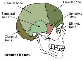
Cranial Bones
- Parietal (2)
- Temporal (2)
- Frontal (1)
- Occipital (1)
- Ethmoid (1)
- Sphenoid (1)
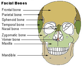
Facial Bones
- Maxilla (2)
- Zygomatic (2)
- Mandible (1)
- Nasal (2)
- Platine (2)
- Inferior nasal concha (2)
- Lacrimal (2)
- Vomer (1)
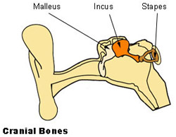
Auditory Ossicles
- Malleus (2)
- Incus (2)
- Stapes (2)
Hyoid (1)
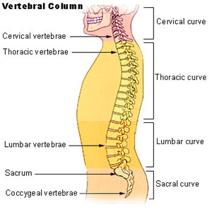
Vetebral Column
- Cervical vertebrae (7)
- Thoracic vertebrae (12)
- Lumbar vertebrae (5)
- Sacrum (1)
- Coccyx (1)
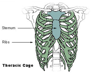
Thoracic Cage
- Sternum (1)
- Ribs (24)
Appendicular Skeleton (126 bones)
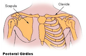
Pectoral girdles
- Clavicle (2)
- Scapula (2)
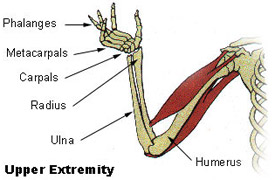
Upper Extremity
- Humerus (2)
- Radius (2)
- Ulna (2)
- Carpals (16)
- Metacarpals (10)
- Phalanges (28)
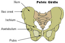
Pelvic Girdle
- Coxal, innominate, or hip bones (2)
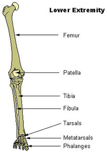
Lower Extremity
- Femur (2)
- Tibia (2)
- Fibula (2)
- Patella (2)
- Tarsals (14)
- Metatarsals (10)
- Phalanges (28)
Articulations
An articulation, or joint, is where two bones come together. In terms of the amount of movement they allow, there are three types of joints: immovable, slightly movable and freely movable.Synarthroses
Synarthroses are immovable joints. The singular form is synarthrosis. In these joints, the bones come in very close contact and are separated only by a thin layer of fibrous connective tissue. The sutures in the skull are examples of immovable joints.Amphiarthroses
Slightly movable joints are called amphiarthroses. The singular form is amphiarthrosis. In this type of joint, the bones are connected by hyaline cartilage or fibrocartilage. The ribs connected to the sternum by costal cartilages are slightly movable joints connected by hyaline cartilage. The symphysis pubis is a slightly movable joint in which there is a fibrocartilage pad between the two bones. The joints between the vertebrae and the intervertebral disks are also of this type.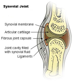
No comments:
Post a Comment